- drparikhrushabh@gmail.com
- +91 8779697851, 9619810512
- Mon - Sat: 10:00am to 6:00pm (Sunday Closed)

Left Atrial Appendage Closure (LAAC)
Meaning:
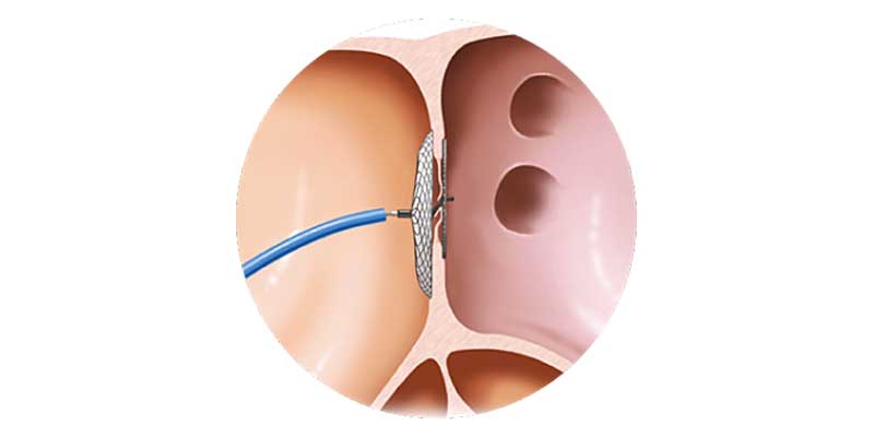
Left Atrial Appendage Closure (LAAC) is a minimally invasive, catheter-based procedure used to reduce the risk of stroke in patients with atrial fibrillation. In atrial fibrillation, the heart beats irregularly, allowing blood clots to form inside a small pouch of the heart called the left atrial appendage. LAAC works by sealing this pouch, preventing clot formation and reducing stroke risk.
Causes:
Atrial fibrillation is the most common heart rhythm disorder and is more frequent in older individuals and those with high blood pressure, diabetes, heart disease, or prior stroke. In atrial fibrillation, about 90% of stroke-causing clots originate from the left atrial appendage, which can allow clots to travel to the brain and block blood flow.
Treatments:
Stroke prevention in atrial fibrillation is usually achieved with blood-thinning medications. However, when long-term anticoagulant use is not suitable due to bleeding risk or frequent falls, LAAC offers an effective alternative. The procedure involves placing a specialized device through a vein in the leg to seal the left atrial appendage under imaging guidance.
Prevention:
Early diagnosis and proper management of atrial fibrillation are essential to prevent stroke. Regular heart rhythm monitoring, appropriate medication use, lifestyle modification, and timely consideration of procedures such as LAAC help reduce long-term complications and improve quality of life.
Frequently Asked Questions
What is atrial fibrillation and how is it related to stroke?
Atrial fibrillation is an irregular heartbeat that can cause blood clots to form in the heart. These clots may travel to the brain and cause a stroke, most commonly originating from the left atrial appendage.
Why is Left Atrial Appendage Closure performed?
LAAC is performed to reduce stroke risk in patients with atrial fibrillation who cannot safely take long-term blood-thinning medications.
When is LAAC recommended?
LAAC is recommended for patients with atrial fibrillation who have a high risk of stroke and an increased risk of bleeding or intolerance to anticoagulant therapy.
What tests are needed before the procedure?
Tests usually include ECG, 2D echocardiography, transesophageal echocardiography (TEE), and sometimes a CT scan to assess heart anatomy and plan the procedure safely.
What happens after the procedure?
Follow-up is typically done after 45 days to confirm complete closure of the appendage. Blood-thinning medication may be stopped if closure is successful, and low-dose aspirin is usually continued long term.